

Scientists have created half-human brain mice — and this is not science fiction. In a study published in the journal Nature on September 16, 2026, a team led by neuroscientist Sergiu Pașca at Stanford University reported that they transplanted lab-grown human brain tissue into specially engineered mice. The human cells did not merely survive: they expanded dramatically, wired themselves into the animals’ nervous systems, and ultimately accounted for the overwhelming majority of the animals’ cerebral cortex. The team calls these hybrid animals “xenocortical” mice, and researchers believe the model could open an entirely new window into human brain development and disorders that are currently almost impossible to study in living tissue.
The announcement immediately captured global attention, with coverage from Reuters, Smithsonian Magazine, Science News, and News-Medical — a sign of just how rare it is for a single neuroscience study to rewrite what researchers thought was possible in a living animal model.
🔬 Key Facts: The Half-Human Brain Mice Study
- Published: September 16, 2026, in the journal Nature
- Led by: Sergiu Pașca, neuroscientist at Stanford University
- The animals: “Apallial” mice — genetically engineered to lack most of their cerebral cortex. As adults, they have only about 2% of the cortical tissue of ordinary mice, leaving a large cavity.
- The transplant: Human cortical organoids, grown for about two months in lab dishes from reprogrammed skin or blood cells of healthy human donors
- Timing: The organoids were surgically placed into the brains of 2-day-old mouse pups
- Survival rate: 86.2% graft survival among 29 mice
- Growth: MRI scans showed a 4.7-fold increase in graft volume between 2 and 3 months after transplantation
- Human share: At 3 months, human-derived tissue accounted for 91.9% of the combined cortical tissue volume
- Integration: Human neurons extended projections, formed synapses, and connected with the mouse’s remaining brain regions (palaeocortex, thalamus, pallidum) and the cervical spinal cord
- Behavior: At 3–6 months after surgery, the xenocortical mice performed broadly similarly to normal mice of the same age, with subtle differences in gait and memory
- Purpose: A new model for studying profound autism, epilepsy, cerebral palsy, and schizophrenia — because living human brain tissue is essentially inaccessible for research for ethical reasons
What Exactly Are Half-Human Brain Mice?
The phrase “half-human brain mice” sounds dramatic, and in a sense it is — but the reality is more precise, and more interesting, than the headline suggests. These are not mice that were born with human brains, nor are they part human in any broad sense. They are laboratory mice whose cerebral cortex — the folded outer layer of the brain involved in perception, memory, and complex thought — is made up overwhelmingly of human cells.
The name the research team uses is “xenocortical” mice: “xeno” from the Greek for foreign or strange, combined with “cortical” for the cortex. The animals began as “apallial” mice, a line of mice genetically engineered to develop without most of their cerebral cortex. As adults, these mice possess only about 2% of the cortical tissue found in ordinary mice, leaving a large cavity where much of the cortex would normally be.
That empty space was the key to everything that followed. In earlier experiments by the same team, human brain tissue transplanted into ordinary animals had to compete for room with the host’s own fully formed cortex. The apallial mice removed that competition entirely: there was simply a large, open cavity waiting to be filled.
When the researchers placed lab-grown human cortical organoids into the brains of these mice at just two days old, the human tissue found an environment where it could expand far beyond anything seen before. Three months later, the graft had grown so extensively that human-derived cells made up 91.9% of the combined cortical tissue volume — the basis for the “half-human” description that has captured the public imagination.
It is worth being clear about what these animals are and are not. They are research models built to answer specific scientific questions. The human cells are neurons and their supporting machinery, grown from donor cells — not a transplanted “mind.” The significance lies not in the headline but in the biology: for the first time, researchers have a living animal in which human cortical tissue develops, connects, and functions at this scale.
The Problem This Research Is Trying to Solve
To understand why this study matters, it helps to understand the wall that brain researchers have been hitting for decades. The human brain is arguably the most complex structure in the known universe, and it is also one of the least accessible. For ethical reasons, scientists cannot experiment on living human brain tissue — you cannot remove samples from a healthy person’s brain, test drugs on it, or watch how it develops in real time.
Researchers have developed workarounds, but each comes with limits. They can study the brains of other animals, yet a mouse or rat brain is not a human brain: the cortex is smaller, simpler, and organized differently. They can grow human neurons and brain organoids in dishes, but a dish cannot reproduce the rich environment of a living body — the blood supply, the sensory input, the connections to the rest of the nervous system.
Transplanting human organoids into animals was an attempt to get the best of both worlds, and the Stanford team had already tried it. In earlier work, they transplanted human cortical organoids into newborn rats and saw something encouraging: the human neurons developed in more advanced ways than they ever had in culture, clearly benefiting from the living environment. But there was a catch. The rat’s own native cortex was still there, occupying the same space and competing with the graft for room to grow and wire up.
The apallial mouse was the team’s answer to that limitation. By starting with an animal that had almost no cortex of its own, the researchers could give the human tissue something it had never had before: space.
How the Stanford Team Built the Half-Human Brain Mice
The experiment unfolded in carefully planned stages, each one building on the last. Here is how the team did it, step by step:
- Start with human cells. The researchers took skin or blood cells from healthy human donors and reprogrammed them into stem cells — cells that can develop into almost any cell type in the body.
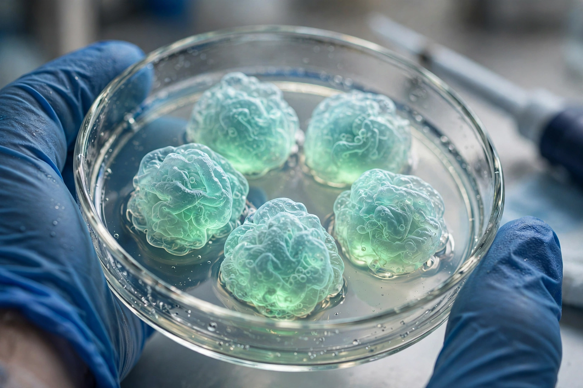
- Grow miniature cortexes. From those stem cells, the team grew human cortical organoids in lab dishes — three-dimensional clusters of developing human brain tissue. The organoids were allowed to mature for about two months.
- Prepare the hosts. In parallel, the team bred apallial mice — animals genetically engineered so that most of their cerebral cortex never develops, leaving a large cavity.
- Perform the transplant. When the mice were just 2-day-old pups, surgeons placed the two-month-old organoids into their brains. Transplanting so early in life meant the graft could grow alongside the developing brain rather than being inserted into a finished one.
- Watch and measure. Over the following months, the team tracked the grafts with MRI scans, examined how the cells connected, recorded their electrical activity, and tested the animals’ behavior.
The numbers tell a striking story. Among 29 mice, the graft survival rate was 86.2% — the great majority of transplants took hold. MRI scans showed a 4.7-fold increase in graft volume between two and three months after transplantation, a remarkable expansion in a short window. And by the three-month mark, human-derived tissue accounted for 91.9% of the combined cortical tissue volume.
Put simply: the human tissue did not just survive in the mouse brain. It thrived there, filling the available space and becoming the dominant tissue of the cortex.
What the Researchers Found
The team did not stop at showing that the grafts survived — they set out to prove that the human tissue was genuinely integrated and functional. The results, published in Nature, were comprehensive.
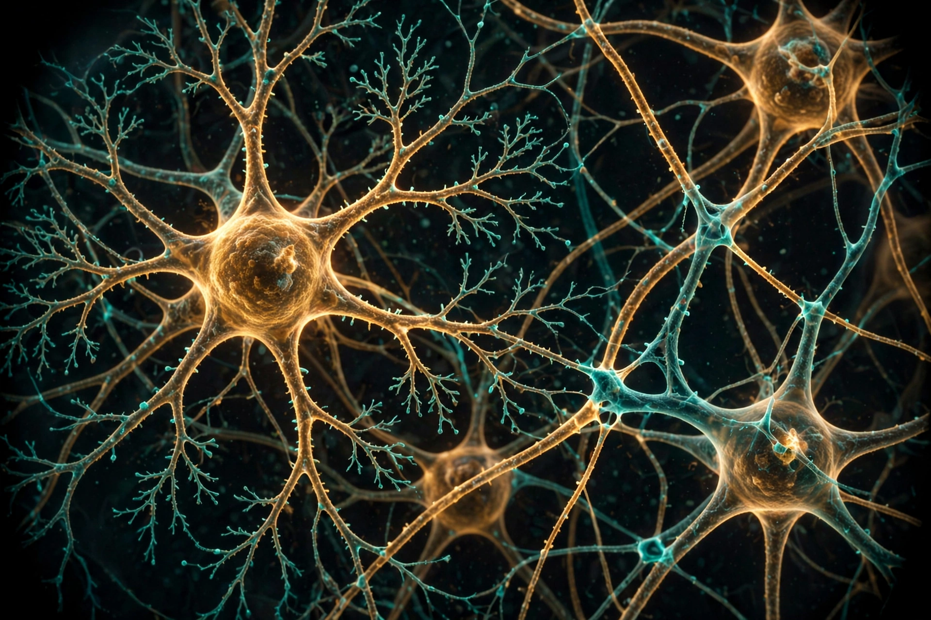
First, the anatomy. The human neurons extended projections — long fibers that reach out to other brain areas — and formed synapses, the junctions through which neurons communicate. Crucially, they integrated with the mouse’s remaining brain architecture: the palaeocortex, the thalamus, and the pallidum, as well as the cervical spinal cord. In other words, the human tissue was not sitting isolated in the cavity. It was wiring itself into the animal’s nervous system, connecting human cells with the host’s surviving brain regions and even reaching down toward the spine.
Second, the activity. Using calcium imaging and local field potential recordings — techniques that reveal when groups of neurons fire together — the researchers observed coherent network-wide bursts. The human neurons were not just present; they were participating in organized, synchronized electrical activity with the rest of the brain. That is a strong indicator that the grafted tissue was functioning as part of a living circuit, not as a passive lump of cells.
Third, the behavior. Between three and six months after surgery, the team put the xenocortical mice through behavioral testing. The headline result: the hybrid animals performed broadly similarly to normal mice of the same age. There were subtle differences — the researchers noted variations in gait and memory — but nothing resembling a wholesale transformation of behavior.
Summing up what the work makes possible, Pașca told Reuters, which covered the study on September 16, 2026:
“This gives us a way to study human neural tissue across several levels, from genes and individual cell types to circuits and functional consequences in an animal. We can begin to ask how disease-associated human genetic changes alter neural development and circuitry and whether potential treatments can prevent or correct those changes,” — Sergiu Pașca (Reuters).
Why Half-Human Brain Mice Could Change Brain Research
The purpose of this work is not spectacle — it is medicine. The team built this model to study human brain development and disorders that have long resisted investigation: profound autism, epilepsy, cerebral palsy, and schizophrenia.
These conditions share a frustrating feature for researchers: their roots lie in the developing human brain, the one organ that cannot be directly studied in living people. Animal models can only approximate human brain development, and cells in a dish cannot show how a disorder plays out across circuits and behavior. A living animal carrying large amounts of functional human cortical tissue could bridge that gap.
The potential applications follow directly from the study’s design. Researchers could introduce disease-associated human genetic changes into the organoids and watch, in a living brain, how those changes alter neural development and circuitry — exactly the kind of experiment Pașca described to Reuters. They could then test whether potential treatments prevent or correct those changes, all within a system where human neurons develop in a living environment rather than a dish.
It is early, and the field is careful not to overpromise. But the direction is clear: a model that lets scientists study human neural tissue across several levels — genes, individual cell types, circuits, and functional consequences in an animal — is something neuroscience has never had. Smithsonian Magazine’s coverage on September 17, 2026 described the hybrid animals as potentially revolutionizing our understanding of neurological disorders — a measure of both the excitement and the expectations now attached to the work.
Where This Story Fits in Today’s Science Events
Science does not happen in a vacuum — breakthroughs land in a culture already buzzing with events, rivalries, and trending stories. The xenocortical mouse study is a reminder that research milestones can become public moments: the kind of story people discuss alongside everything else capturing the world’s attention, from championship clashes like the Chivas–América Clásico Nacional, one of the biggest events in Mexican football, to the awareness days and celebrations we cover here on Events Day.
What makes this story travel so far beyond specialist journals is the combination of genuine scientific substance and a genuinely startling image: human brain tissue, growing and wiring itself inside a living mouse. That is why outlets from Reuters to Smithsonian Magazine to Science News all covered it within days of publication — and why it is likely to remain a reference point in debates about brain research for years to come.
The Ethics Questions Still Without Answers
Any experiment that puts human brain tissue into an animal raises questions that go beyond the laboratory — and this study, with its unprecedented scale, raises them more sharply than ever. It is important to be honest about this: these questions do not yet have settled answers.
The first open question concerns long-term behavioral outcomes. The published testing covered three to six months after surgery and found the xenocortical mice broadly similar to normal mice, with only subtle differences in gait and memory. But what happens over a longer lifespan? The study does not answer that, and the team has not claimed otherwise. How these animals develop, age, and behave over time remains genuinely unknown, and longer-term observation will be needed before anyone can speak with confidence.
The second open question is about ethical review frameworks. As human neural tissue makes up a larger and larger share of an animal’s brain — 91.9% of the cortical volume in this study — the existing guidelines for animal research and for human tissue research start to strain. When does a brain graft cross a line that current rules were not written for? There is no consensus answer, and ethicists, scientists, and institutions will need to build those frameworks together, in the open.
The third open question is about regulatory implications. If models like this become standard tools for disease research and drug testing, which agencies oversee them, and under what rules? The science is moving faster than the rulebooks — a familiar pattern in biotechnology — and one that calls for deliberate public conversation now rather than after-the-fact scrambling later.
None of this diminishes the study’s scientific achievement. But the researchers themselves frame the work as a tool for understanding disease, not as an endpoint. The ethical conversation will need to keep pace with the biology, and it is only just beginning. News-Medical’s detailed report on the study walks through the findings for readers who want the full technical picture.

At a Glance: Key Details of the Xenocortical Mouse Study
| Detail | What the study reported |
|---|---|
| Publication | Nature, September 16, 2026 (Wednesday) |
| Senior author | Sergiu Pașca, neuroscientist at Stanford University |
| Animal model | “Apallial” mice, genetically engineered to lack most of their cerebral cortex (about 2% of normal cortical tissue as adults) |
| Donor cells | Skin or blood cells from healthy human donors, reprogrammed into stem cells that can become almost any cell type |
| Organoid age at transplant | About 2 months old |
| Recipient age | 2-day-old mouse pups |
| Graft survival | 86.2% among 29 mice |
| Volume growth | 4.7-fold increase in graft volume between 2 and 3 months (MRI) |
| Human tissue share | 91.9% of combined cortical tissue volume at 3 months |
| Integration | Human neurons extended projections, formed synapses, and integrated with the palaeocortex, thalamus, pallidum, and cervical spinal cord |
| Neural activity | Calcium imaging and local field potential recordings showed coherent network-wide bursts |
| Behavior | 3–6 months after surgery: broadly similar to normal mice of the same age, with subtle differences in gait and memory |
| Target disorders | Profound autism, epilepsy, cerebral palsy, schizophrenia |
| Earlier work | Human cortical organoids transplanted into newborn rats showed more advanced neuronal development than in culture, but the rat’s native cortex competed for space |
Frequently Asked Questions About Half-Human Brain Mice
What are half-human brain mice?
They are laboratory mice whose cerebral cortex is made up overwhelmingly of human cells. Stanford researchers transplanted lab-grown human cortical organoids into “apallial” mice — animals engineered to develop with only about 2% of normal cortical tissue — and the human grafts expanded until they accounted for 91.9% of the combined cortical volume. The team calls them “xenocortical” mice.
Who created them, and where was the study published?
The study was led by neuroscientist Sergiu Pașca at Stanford University and published in the journal Nature on September 16, 2026. It was covered the same week by Reuters, Smithsonian Magazine, Science News, and News-Medical.
How did the scientists make the xenocortical mice?
They reprogrammed skin or blood cells from healthy human donors into stem cells, grew human cortical organoids in lab dishes for about two months, and then surgically placed the organoids into the brains of 2-day-old apallial mouse pups. Among 29 mice, the graft survival rate was 86.2%, and MRI scans showed a 4.7-fold increase in graft volume between two and three months after transplantation.
Did the human brain tissue actually work inside the mice?
Yes, according to the study’s measurements. The human neurons extended projections, formed synapses, and integrated with the mouse’s remaining brain regions — the palaeocortex, thalamus, and pallidum — as well as the cervical spinal cord. Calcium imaging and local field potential recordings revealed coherent network-wide bursts, indicating that the human cells were participating in organized, synchronized brain activity.
Did the mice behave normally?
Broadly, yes. In behavioral testing conducted three to six months after surgery, the xenocortical mice performed similarly to normal mice of the same age. The researchers did note subtle differences in gait and memory, but nothing resembling a fundamental change in behavior.
What diseases could this research help scientists understand?
The model was built to study human brain development and disorders that are difficult to investigate because living human brain tissue is essentially inaccessible for ethical reasons: profound autism, epilepsy, cerebral palsy, and schizophrenia. Researchers hope to use it to see how disease-associated human genetic changes alter neural development and circuitry, and to test whether potential treatments can prevent or correct those changes.
Does this mean the mice are part human or conscious like people?
There is no evidence for that, and the researchers do not make that claim. The human cells are neurons grown from donor cells, integrated into a mouse’s nervous system — the team describes the animals as research models for studying disease. Long-term behavioral outcomes, ethical review frameworks, and regulatory implications remain open questions that the scientific community has yet to resolve.
📅 More from Events Day
- Dolly Parton Day — explore our coverage of the celebration honoring the country music legend.
- Suicide Prevention Awareness Month — learn more about this important month of awareness, support, and hope.
The Bottom Line on Half-Human Brain Mice
In a single study, Stanford researchers turned one of brain science’s longest-standing limitations into an opportunity. By giving human cortical organoids the one thing they had never had — room to grow inside a living brain — the team achieved an 86.2% graft survival rate, a 4.7-fold expansion in graft volume, and a cortex that was 91.9% human-derived within three months. The human neurons wired into the mouse’s surviving brain regions and the spinal cord, fired in coherent networks, and left the animals behaving broadly like ordinary mice.
What comes next is just as important as what has been shown. Longer-term studies will need to track how these animals develop over their full lifespans. Disease-modeling experiments will test whether the xenocortical mouse can truly illuminate profound autism, epilepsy, cerebral palsy, and schizophrenia. And the ethical and regulatory conversations — about review frameworks, oversight, and where the lines should be drawn — will need to keep pace with the biology.
For now, the half-human brain mice stand as one of the most striking neuroscience achievements of 2026: a living model of the human cortex that scientists can finally study in action. We will keep following this story as it develops.